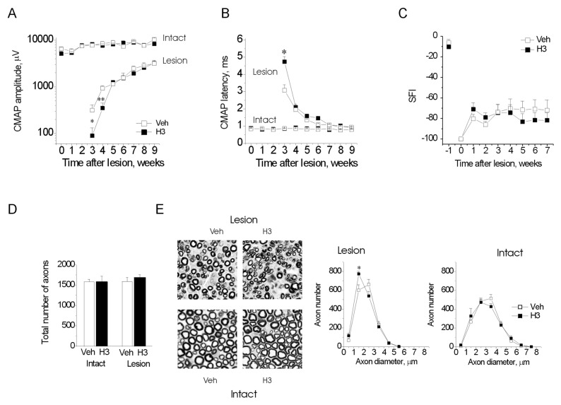
Figure 2.
H3 delays axonal regeneration after nerve transection in mice. (A, B) Development of CMAP amplitude (A) and conduction velocity (B) in unoperated (Intact) and operated (Lesion) nerves in the vehicle (Veh)- and H3-treated animals. (C) Recovery of the functional index (SFI). (D, E) Total number of fibers (D) and myelinated fiber distributions (E, right) at 8 wks after lesion. (E, left) Representative images of regenerated sciatic nerves from the vehicle- and H3-treated animals at 8 wks after transection, 50 × 50 μm fields are shown. (A–E) *p < 0.05 versus Veh, two-tailed t test. (A–C) Number of animals, n = 7/7 (Veh/H3). (D, E) n = 4/4.

