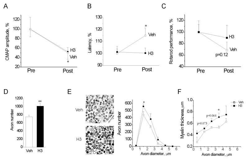
Figure 5.
The H3 peptide delays neurodegeneration caused by deficiency in the myelin P0 gene. (A–C) Development of CMAP amplitude (A), conduction velocity (B) and rotarod performance (C) in P0−/− mice treated with vehicle (Veh) or the H3 peptide for 30 d. Measurements were performed before treatment (Pre) start and immediately after treatment finish (Post) and were normalized to the Pre value in each group (set to 100%). *p < 0.05 versus Pre, two-tailed t test. (D–F) Total number of fibers (D), myelinated fiber distribution (E) and myelin thickness distribution (F) at the end of the treatment period. (E, left) Representative images of sciatic nerves from the Veh- and H3-treated animals at the end of the treatment period; 50 × 50 μm fields are shown. *p < 0.05 versus Veh, two-tailed t test. (A–C) Number of analyzed nerves, n = 13/15 (Veh/H3). (D, E) n = 6/8.

