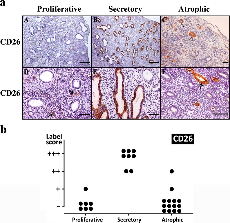
Fig. 6.
a Immunolocalization of CD26 in proliferative (a, d), secretory (b, e) and atrophic (c, f) endometria. CD26 was immunodetected in the blood vessels (arrows) and in glandular epithelia of secretory endometrium (b, e). Note that only a few isolated glands are labeled in atrophic endometrium (c, empty arrowheads). Scale bars = 500 μm (a, b) and 100 μm (c–f). b Label intensity score of CD26 in the glandular epithelium of proliferative, secretory and atrophic endometria. Maximal score is found in secretory endometria

