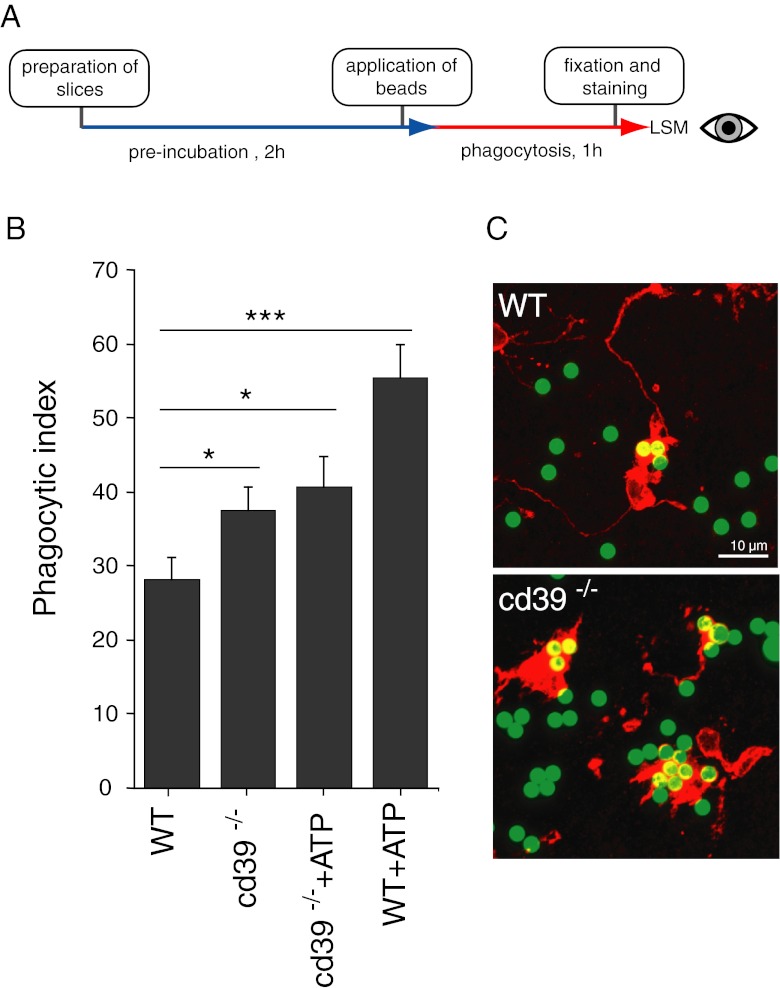
Fig. 2.
a Flow diagram of the in situ phagocytosis assay using acute brain slices. b Microglial phagocytosis was higher in cd39−/− slices compared to slices from wild-type adult mice. Application of 100 μM ATP led to increased phagocytosis in WT slices but had no effect in cd39−/− slices. Data presented as mean ± standard error of the mean. c Representative examples of fluorescent beads (green) phagocytosed by microglia (red) in acute brain slices from wild-type and cd39−/− mice, scale bar 10 μm. Significant p values are as follows: single asterisk indicates p < 0.05; double asterisks indicate p < 0.01; triple asterisks indicate p < 0.001

