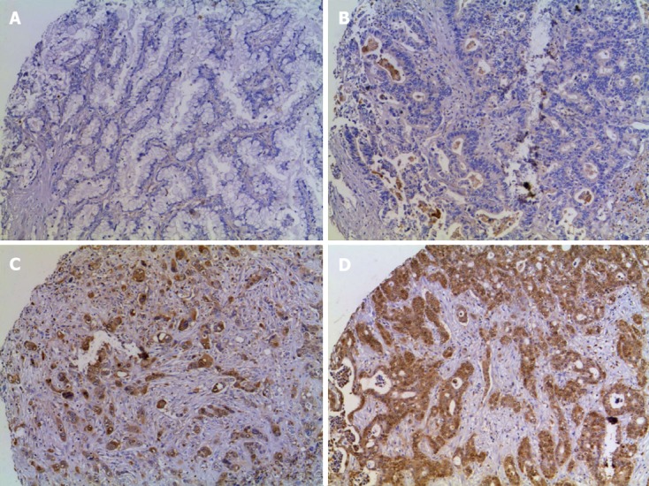
Figure 1.

Immunostaining of annexin A1 protein expression in cholangiocarcinoma tissue microarrays. A-D: Tissue microarray of cholangiocarcinoma samples was stained with anti-annexin A1 antibody and counter stained with Mayer's hematoxylin and represented by A for negative, B for +, C for ++, and D for +++ when expression was < 10%, 10%-25%, 26%-75% and > 75%, respectively (magnification, × 200).
