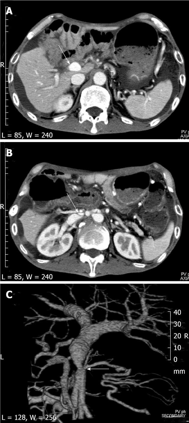
Figure 1.

Computed tomography showed short segmental stenosis of the portal vein in the region of the anastomosis, severe ascites, and liver atrophy. A, B: Computed tomography (CT) scan shows severe ascites and liver atrophy (arrow) (A), and stenosis of the portal vein (arrow) behind the superior mesenteric artery (B); C: The image of the 3D reconstruction of the portal vein shows short segmental stenosis in the region of the anastomosis (arrow).
