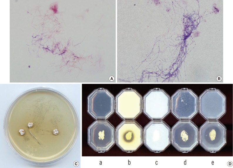
Fig. 2.
(A) The Gram-stained smear of a cultured colony reveals thin and filamentous branching rods (×1,000). (B) Modified-Kinyoun staining reveals weakly positive dotted rods (×1,000). (C) The morphology of colonies of the isolate on SDA after 3-week incubation. (D) Biochemical tests, including the opacification of Middlebrook 7H10 (a) and the hydrolysis of casein (b), xanthine (c), hypoxanthine (d), and tyrosine (e), were interpreted after a 5-day incubation at 35℃. The organism was negative for Middlebrook 7H10 opacification and xanthine hydrolysis but positive for casein, hypoxanthine, and tyrosine hydrolysis (lower row). The un-inoculated media (upper row) are shown for comparison.
Abbreviation: SDA, Sabouraud dextrose agar.

