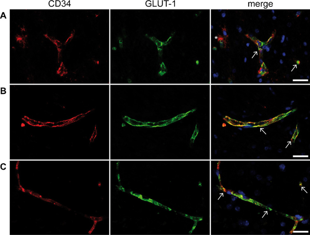
Fig. A4.
Fluorescent double-immunohistochemistry of GLUT-1 and CD34. Combined images reveal a co-localization of GLUT-1 (green) immunoreactivity and CD34 (red) positive vascular endothelial cells (white arrows) in lesional (A) and perilesional (B) region of ganglioglioma WHO grade I and lesional region of diffuse astrocytoma WHO grade II (C). Characteristic satellite cells in the tumor region of gangliogliomas are strongly CD34 positive (asterisk in A). Microscopically no differences of GLUT-1 expression between the analyzed entities were observed. Scale bar = 50 µm. (For interpretation of the references to color in this figure legend, the reader is referred to the web version of the article).

