
Figure 2. Histopathological study.
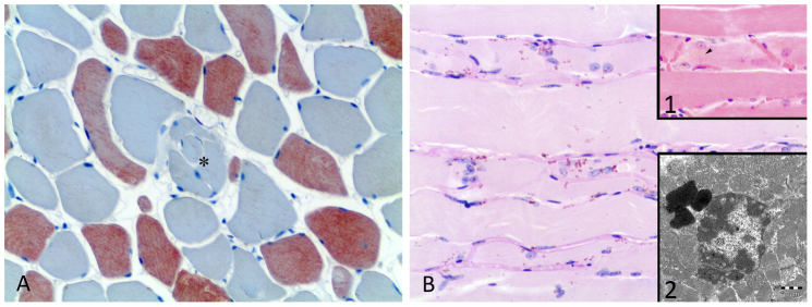
(A): Cross section of a muscle from a senile spotted dolphin specimen. The muscle exhibits a variety of abnormal features consisted of an increased variations in the fibre sizes and longitudinal splitting (*). Immunostaining for the fast MHC isoform. 20×. (B): Longitudinal section of a muscle from a senile spotted dolphin specimen. The muscle exhibits a large amount of juxtanuclear lipofuscin and significant vascularisation within the fibres. PAS. 20×. 1: Lipofuscin appears as a faint yellow pigment, mostly adjacent to the nucleus. HE. 20×. 2: An EM image showing the lipofuscin accumulation was located mostly in the mitochondria-rich subsarcolemmal area. Bar = 1 μm.