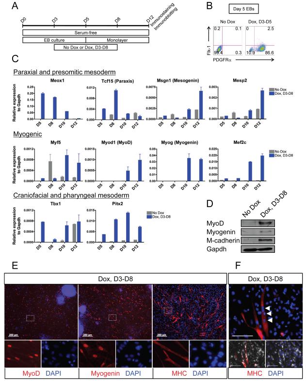
Figure 5. Mesp1 promotes paraxial mesoderm and myogenic derivatives in the absence of serum-derived factors.
(A) Scheme depicting the protocol used to examine the effect of Mesp1 on ES differentiation in serum-free conditions.
(B) FACS analysis of mesoderm markers Flk-1 and PDGFRα in day 5 EBs grown under serum-free conditions.
(C) Quantitative RT-PCR for various lineage-specific markers in serum-free culture conditions in which Mesp1 was induced from day 3 to day 8 (blue bars) or not induced (gray bars) (n=3).
(D) Immunoblot showing upregulation of myogenic proteins upon Mesp1 induction from day 3 to day 8 under serum-free conditions.
(E) Immunostaining for myogenic markers, MyoD (left), Myogenin (middle) and MHC (right), in EB-derived cells induced by Mesp1 from day 3-8 cultured in the absence of serum. Areas depicted by the white dotted rectangle are magnified to demonstrate the localization of the nucleus (DAPI) and the nuclear (MyoD and Myogenin) or cytoplasmic (MHC) markers of interest (bottom panels). No MyoD+, Myogenin+ or MHC+ cells were observed in control (No Dox) wells (not shown). Bar = 200 μm
(F) Immunostaining showing the presence of multiple nuclei (white arrowheads) in Mesp1-induced ES cell-derived MHC+ myotube. The red channel (MHC) and the blue channel (DAPI) are individually merged with the phase contrast channel to indicate that multiple DAPI+ nuclei are present in the same MHC+ myogenic cell (bottom panels). Bar = 100 μm
Mean ± SEM is shown in panel C.

