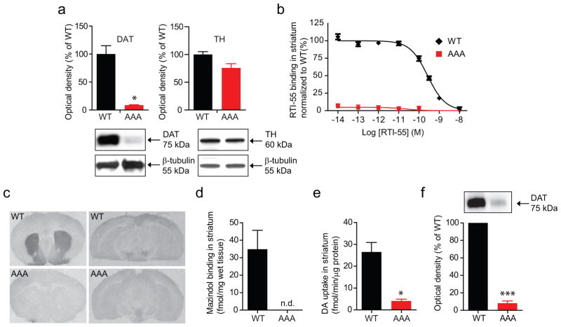
Figure 2. Assessment of DAT binding sites and dopamine transport in DAT-AAA mice.
(a) Immunoblotting confirmed a major loss of DAT expression in striatum while TH levels were similar to WT. Upper panels, densitometric analysis of immunoblots for WT and DAT-AAA mice, *P<0.05, non-parametric Mann-Whitney test (means ± s.e.m., n=4). Lower panels, representative immunoblots for DAT (left) and TH (right) in striatal lysates from WT and DAT-AAA mice. (b) Competition binding experiments on striatal membranes using the high-affinity DAT ligand [125I]-RTI-55. Data are normalized to WT littermate controls (n=4). [125I]-RTI-55 binding was analysed by non-linear regression analysis using Prism 5.0. The affinity constants (KI) were determined assuming one-site binding. Bmax and Kd values were obtained from the IC50 value estimated by assuming a sigmoidal one-site curve (One site–Fit LogIC50). Bmax: WT, 290 ± 50 fmol/μg protein; DAT-AAA, 30 ± 6.2 fmol/μg protein (means ± s.e.m., n=4); Kd: WT, 0.127 [0.105 – 0.154] nM; DAT-AAA, 0.334 [0.108 – 1.062] nM (means [s.e.m. interval], n=4). (c, d) [3H]-mazindol autoradiography shows a high density of DAT binding sites in striatum from WT mice, while binding is absent in DAT-AAA mice (means ± s.e.m., n=4). Midbrain region shows lower density of binding sites in WT and binding is practically absent in DAT-AAA mice. (e) Extensive loss of [3H]-dopamine uptake (0.125 μM) in striatal synaptosomes from DAT-AAA mice relative to WT, *P<0.05, non-parametric Mann-Whitney test (means ± s.e.m., n=4–5). (f) Surface biotinylation of striatal slices show a dramatic loss of surface expressed DAT in DAT-AAA mice (% of WT control ± s.e.m., one-sample t test, n=3, *** P<0.001).

