
Figure 5. Dopaminergic neurons show reduced DAT-AAA surface expression and no ER retention of DAT-AAA.
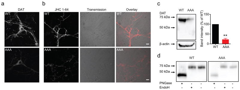
(a) Visualization of DAT in dopaminergic neurons from WT and DAT-AAA mice by immunostaining and confocal microscopy. DAT-AAA labelling intensity was dramatically reduced compared to WT. Labelling was most intense in somas, but DAT-AAA was clearly present in extensions as well. Images are representative of stainings of three independently prepared cultures (b) Confocal live imaging of midbrain neurons derived from WT or DAT-AAA mice. Neurons were stained with the fluorescent cocaine analogue, JHC 1-64 (10 nM), to obtain specific labelling of endogenously expressed WT and DAT-AAA present in the plasma membrane. Neurons from DAT-AAA mice exhibited markedly reduced surface labelling. Pictures shown are representative of labelling of three independently prepared cultures. Scale bars=10 μm. (c) Immunoblotting of membrane fractions derived from whole brains of new born pups (P1–P3). Left panel, representative immunoblot. A single DAT band (~70kDa) is detected in membrane fractions from both WT and DAT-AAA pups, albeit with markedly reduced intensity for DAT-AAA. Note that no immature DAT bands were detected in either phenotype. Right panel, densitometric analysis of immunoblots shown as % of WT, means ± s.e.m., **P<0.01, one sample t-test. (d) EndoH and PNGase F treatment of neuronal lysates. Immunoblotting of lysates from cultured dopaminergic neurons likewise show that untreated lysates give rise to a single DAT band (~70 kDa), for both WT and DAT-AAA, with no detectable immature bands. Both the WT and the DAT-AAA bands are insensitive to Endo H treatment, but elutes at ~45 kDa after deglycosylation by PNGase F (n=3).