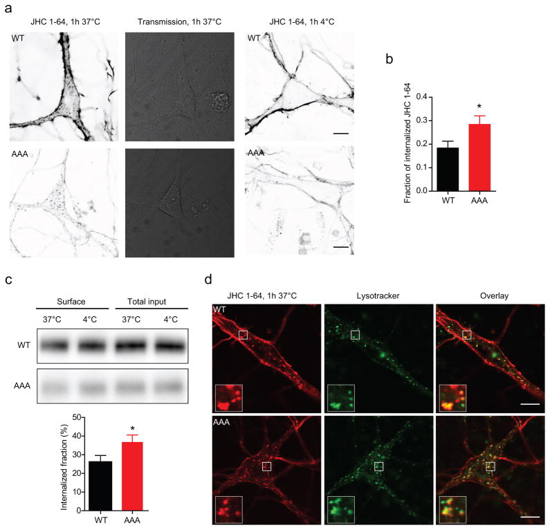
Figure 6. Visualization of constitutive DAT internalization in WT and DAT-AAA neurons.
(a) Visualization of constitutive internalization of DAT in WT and DAT-AAA neurons by confocal live imaging using the fluorescent cocaine analogue, JHC 1-64. Left panels, Surface expressed DAT was labelled by incubation of neurons with 10 nM JHC 1-64 at 4°C, followed by 1h incubation at 37°C to allow internalization. Middle panels, transmission image. Right panels, 4°C temperature control for internalization. Fluorescence is shown on a grey scale with the highest fluorescence represented by the darkest pixels. Internalization of DAT/JHC 1-64 complexes is seen as JHC 1-64 positive vesicular structures in the neuronal soma and proximal extensions. Similar JHC 1-64 positive vesicles did not appear under 4°C control conditions. Images shown were taken with identical settings to visualize the JHC 1-64 intensity under identical conditions for WT and DAT-AAA neurons. (b) Quantification internalized DAT. The amount of internalized DAT was quantified relative to the total amount of JHC 1-64 labelled DAT and showed that a significantly larger fraction of DAT was internalized in DAT-AAA neurons (n=20 neurons) compared to WT neurons (n= 30 neurons), means ± s.e.m., * P<0.05, non-parametric Mann-Whitney test. (c) Surface biotinylation of striatal slices. Striatal slices were biotinylated after 1h incubation at either 37°C to allow internalization, or at 4°C a non-trafficking permissive temperature control. Upper and middle panels, representative blots of WT and DAT-AAA lysates respectively. Lysates from DAT-AAA and WT mice were run on separate gels to allow better visualization of the DAT-AAA band without reaching saturating levels of the WT band intensity. Lover panel, quantification of internalized fractions i.e. the reduction in surface levels. The internalized fraction was significantly larger in DAT-AAA slices, means ± s.e.m, * P<0.05, one-tailed t-test, n=3. (d) Colocalization between constitutively internalized JHC 1-64/DAT complexes and LysoTracker. A considerable amount of colocalization with LysoTracker is seen for both WT DAT and DAT-AAA. Scale bars=10 μm.

