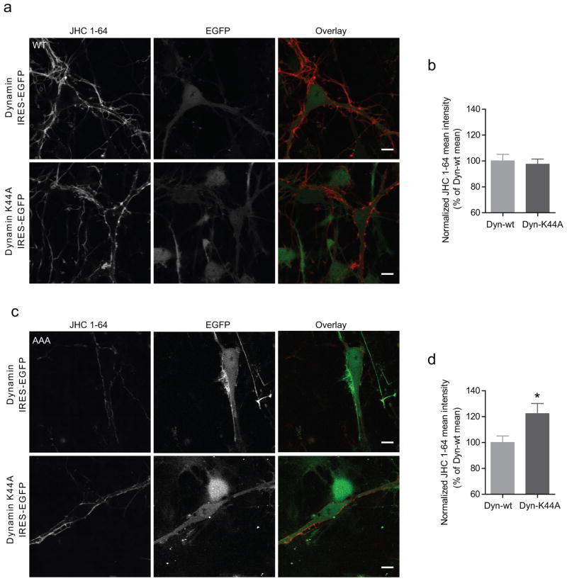
Figure 7. Inhibition of dynamin-dependent internalization increases surface levels of DAT-AAA, but not WT.
(a) Midbrain dopaminergic neurons, derived from WT pups, were transduced at day 1–3 in vitro with lentivirus encoding either WT dynamin or the dominant-negative K44A dynamin mutant, both coupled to EGFP expression. Left panels, JHC 1-64 labelling (10 nM) of surface expressed DAT, performed 8–10 days after transduction, to evaluate the effect of inhibiting DAT internalization on surface expression. Middle panels, EGFP signal. Right panels, overlay of channels. (b) Quantification of JHC 1-64 fluorescence revealed no difference between WT and K44A dynamin transduced WT neurons, means ± s.e.m. (n=34 and 38 neurons for WT dynamin and dynamin-K44A respectively). (c) Midbrain dopaminergic neurons, derived from DAT-AAA pups, transduced with lentivirus encoding either WT dynamin or the dominant-negative K44A dynamin mutant. Left panels, JHC 1-64 labelling (10 nM) of surface expressed DAT. Middle panels, EGFP signal. Right panels, overlay of channels. (d) Inhibition of dynamin-dependent internalization, by dynamin K44A, in DAT-AAA neurons led to a significant increase in the mean JHC 1-64 intensity, relative to control neurons, transduced with WT dynamin (n=25 and 26 neurons for dynamin WT and K44A respectively, * P<0.05, non-parametric Mann-Whitney test). Images shown originate from at least 5 independent stainings from at least 4 independent neuronal preparations. All images of a given genotype have been taken with identical settings for JHC 1-64 detection. Scale bars=10 μm.

