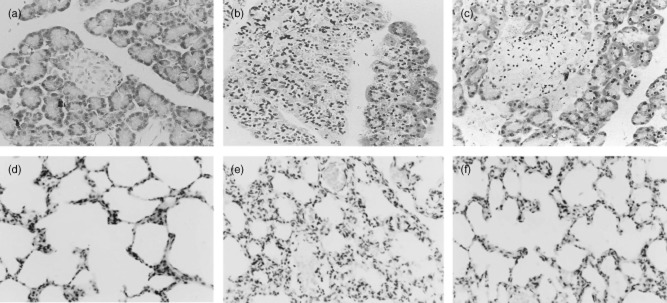
Fig. 2.

Morphological changes of pancreas and lung stained with haematoxylin and eosin (H&E). (a) No histological alteration of pancreas was observed in the pancreas collected from the control group. (b) Histological examination [at 24 h after severe acute pancreatitis (SAP)] of pancreatic sections from pancreatitic rats revealed oedema and acinar cell necrosis as well as inflammatory cells infiltration and an important alteration of the pancreas was also present. (c) Significantly less histological alteration of the pancreas tissue was observed in pancreatitic rats, which received ethyl pyruvate (EP) treatment. Representative H&E-stained section of lung was examined by light microscopy in control rats (d) and in pancreatitic rats (e), and in pancreatitic rats, which received EP treatment (f). Original magnification: ×400. Figure is representative of at least three experiments performed on different experimental days.
