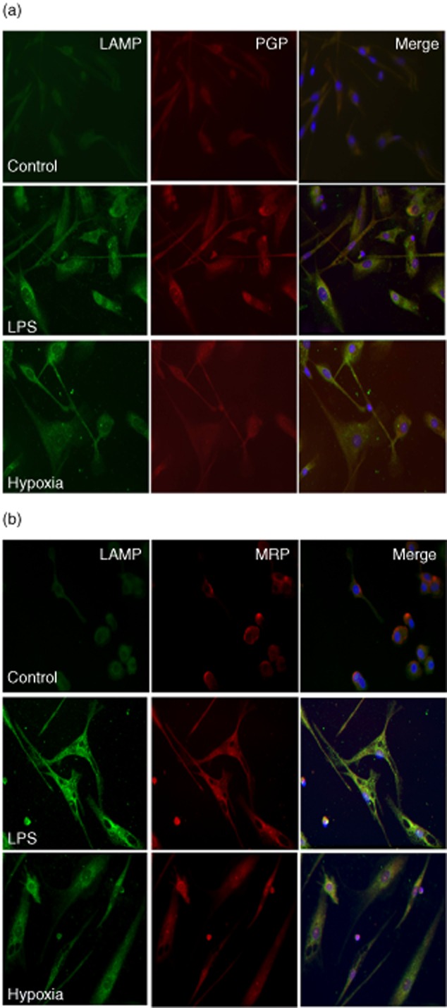
Fig. 2.

Over-expression of P-glycoprotein (Pgp) and multi-drug resistance protein 1 (MRP1) by immunofluorescence in dendritic cells (DCs) after hypoxia and lipopolysaccharide (LPS). Two-colour immunofluorescence images were obtained from DC lysosomal-associated membrane protein (LAMP) antibody [first panel of each column (1), green] and P glycoprotein antibody (JSB1) Pgp (a) or MRP1 (b) antibody [second panel of each column (2), red]. The two images were merged and double-positive cells are shown in the third panel of each row (3). iDCs appear in the first row, LPS-DC in the second row and hypoxia-DCs in the third row.
