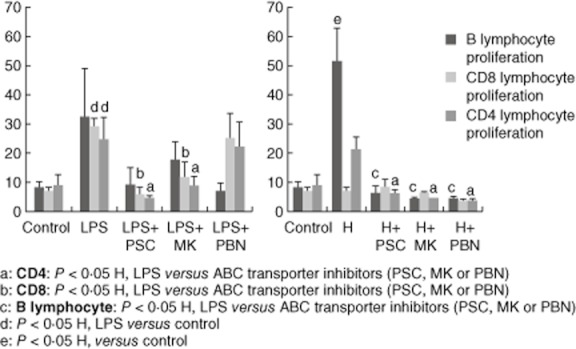
Fig. 6.

T cell proliferation in the mixed lymphocyte reaction (MLR). Lymphocytes were stained with carboxyfluorescein diacetate succinimidyl ester (CFSE) and exposed to mature dendritic cells (DCs) [under hypoxia or lipopolysaccharide (LPS) stimuli] with or without adenosine 5′-triphosphate-binding cassette (ABC) transporter inhibitors. Cell proliferation was determined by flow cytometry after labelling with CD20, CD4 and CD8 antibodies. The results are representative of six independent experiments and expressed as the mean ± standard error.
