
Fig. 3.
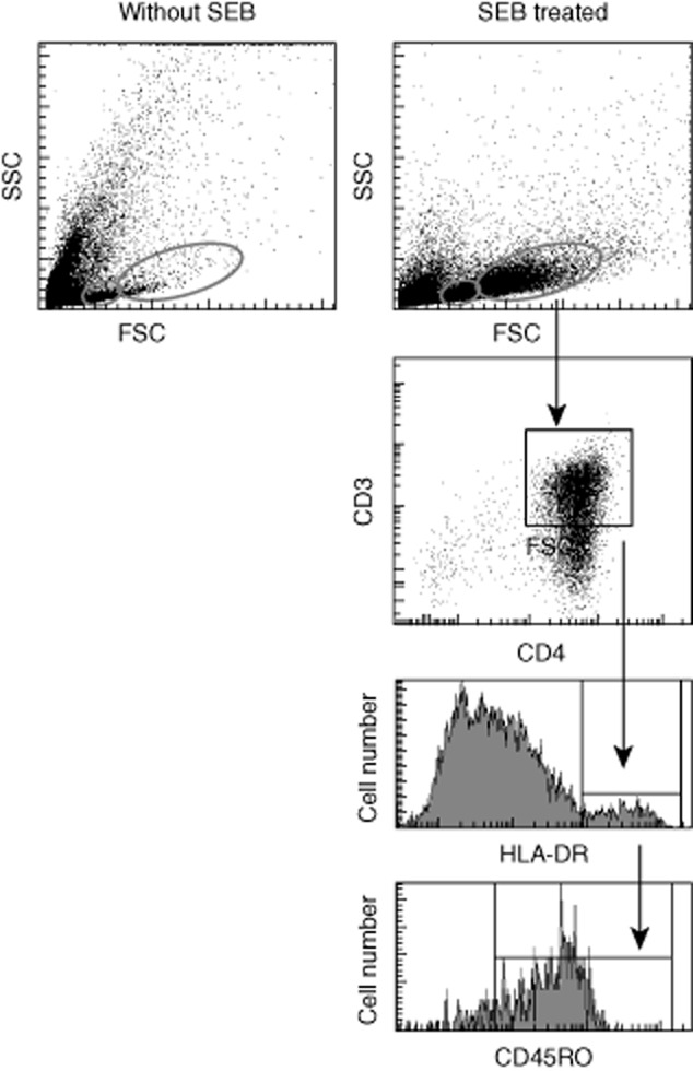
Illustration of gating strategies for analysis of lymphoblasts. Enriched human CD4+ T cells and CD14+ monocytes were incubated separately with medium alone or with one of the chemotherapeutic drugs overnight. The next day, the treated CD14+ and CD4+ cells were mixed with staphylococcal enterotoxin B (SEB) 5 μg/ml and incubated for the time-period indicated for flow cytometric measurement of lymphoblast development. Small non-granular lymphocytes (small red circles) and large granular lymphoblasts (big red circles) were identified on dot-plots, and then large granular lymphoblasts were further analysed based on CD3 versus CD4 dot-plots. Double positive cells were analysed further on a histogram to identify human leucocyte antigen D-related (HLA-DR+) cells, and these cells were analysed further to identify CD45RO-positive cells on a histogram. The cells CD3+, CD4+, Cd45RO+ and HLA-DR+ were considered lymphoblastic.