
Fig. 6.
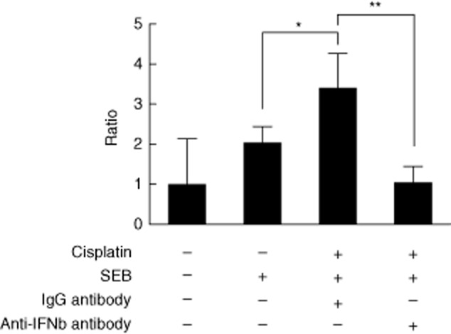
Attenuation of cisplatin-enhanced T cell proliferation by neutralizing interferon (IFN)-β. Enriched human CD4+ T cells and CD14+ monocytes were incubated with medium and cisplatin, respectively, overnight. On the next day the treated 1·8 × 105 CD14+ and 3·6 × 105 CD4+ cells were mixed in a final volume of 1 ml after twice washing. Interferon-β-neutralizing antibody and its immunoglobulin (Ig)G isotype control were added separately to the cell mixture. After 1 h, staphylococcal enterotoxin B (SEB) 5 μg/ml was added and then the mixture was incubated for 5 days for analysis by flow cytometry. The mixed samples without treatment were considered as control. The proliferation ratio of lymphoblasts was calculated for different treatment groups, and then normalized to the value of the control group, which was set to 1. Data shown are the mean values for three donors. Error bars illustrate standard deviation. Significance comparing the groups is indicated (*P < 0·05; **P < 0·01).