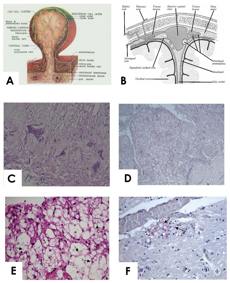
Figure1.
A. Diagram of the structure of human arachnoid granulation (from [10], with permission). B. Diagram illustrating position of the arachnoid granulations between the sub arachnoid space and venous sinus. C. High power view of normal arachnoid granulation from a patient dying of unrelated cause (H&E stain). D and E (high power). Case 1. Arachnoid granulation with large numbers of cryptococcal cells (arrowed) and little inflammation. Mucicarmine stain. F. Case 1. Brain tissue showing cryptococcal cells (arrowed) invading brain adjacent to a blood vessel. Mucicarmine stain. Note the paucity of organisms in the brain tissue compared to the arachnoid granulations.

