Figure 4.
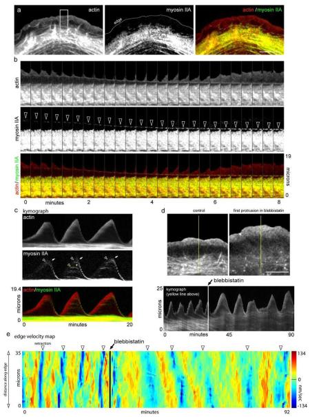
Myosin II activity condenses the lamellipodium into an actin arc. a) Organization of actin-mRFP, Myosin IIA-GFP, and overlay during edge protrusion. Notice myosin II localizes with older actin arcs in the lamella. b) Time-lapse montages of actin-mRFP, myosin IIA-GFP and an overlay showing the co-localization of myosin II with newly forming actin arcs. Arrowheads show co-translocation of myosin IIA and the newly formed actin arc. c) Kymograph showing myosin IIA dynamics over three protrusion/retraction cycles. Arrowheads denote the appearance and arrows denote the movement of myosin IIA. d) Actin-mRFP before and after 25 μm blebbistatin. Kymograph shows the protrusion retraction cycle of the edge before and after blebbistatin. Note that the structure and movement of the actin arcs is diminished in the presence of blebbistatin. e) Protrusion/retraction map showing edge motion before and after blebbistatin addition (arrow). Edge retractions denoted by arrowheads. Scale bars, 10 μm.

