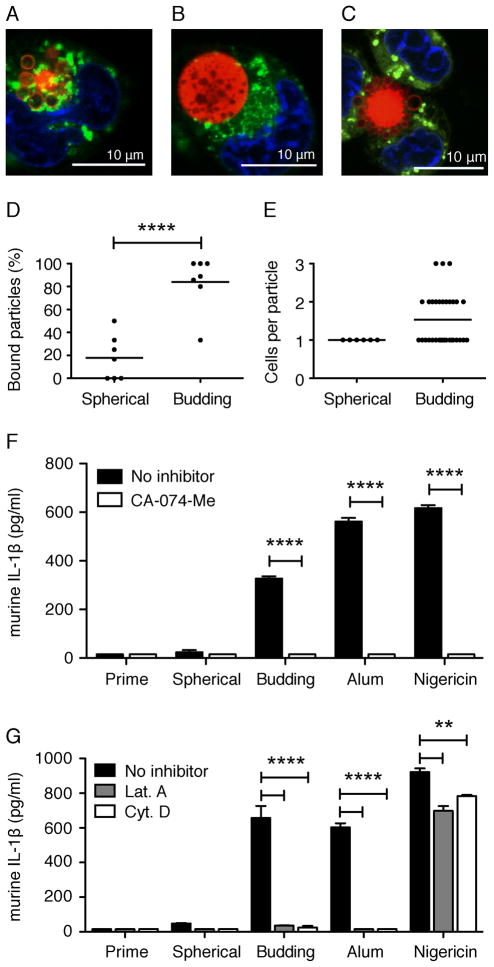
Figure 6. Particle phagocytosis: Budding particles associate with more macrophages than spherical particles.
Confocal microscopy images of macrophage-associated budding (A,C) and spherical (B) particles following a 3 h prime with LPS and 6 h incubation with particles. Lysosomes were visualized with LysoTracker Green. Nuclei were visualized with Hoechst 34580 (blue). Scale bars = 10 μm. Images were taken with a x63 objective. Percent of and average (line) particles bound to macrophages per field of view (D). Total and average (indicated by line) number of macrophages associated with a single particle per field of view (E). Analysis was performed on seven independent fields of view per particle type. Secreted IL-1β from WT immortalized macrophages stimulated for 6 h (F) or 18 h (G) with 100μg budding or spherical particles, 130 μg/ml alum, or 5 μM nigericin in the presence (white bars) or absence (black bars) of 50 μM CA-074-Me (F), 250 nM Latrunculin A (Lat. A), or 1 μM Cytochalasin D (Cyt. D) (G). Cytokine levels are reported as mean + SEM and are representative of two independent experiments performed in duplicate. Significance values are shown as budding versus spherical particles (D) or untreated versus treated cells (F, G). ****, P < 0.0001; **, P< 0.01.

