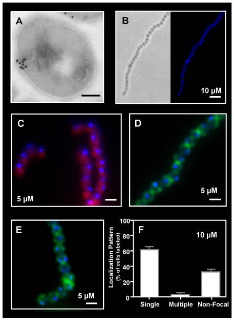
Figure 1. Focal binding of Polymyxin B to the S. pyogenes surface.
The distribution of polymyxin B on the surface of S. pyogenes SF370 following sub-lethal challenge was revealed: (A) by treatment with biotinylated polymyxin B (1:10,000) and immunogold electron microscopy using staining with a streptavidin-gold conjugate (scale bar = 200nm) and (B, C, D, E) by fluorescent microscopy following challenge with dansyl-polymyxin B alone (B) at the concentration indicated in the Figure (scale bar = 1μm) or in cells counterstained with Nile Red (C), fluorescent vancomycin (D) or wheat germ agglutinin Alexa Fluor 488 conjugate (E). Staining patterns following challenge with dansyl-polymyxin B were quantitated as described in the Experimental Procedures (F). Data represents the mean and standard error of the mean (SEM) derived from at least 3 independent experiments and examination of a minimum of 1000 stained cells. The number of cells with a single focus was significantly higher than any other staining pattern (P < 0.0001).

