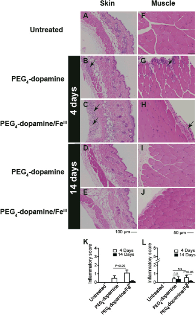
Figure 6.
Tissue reaction to 100 µl of cross-linked and un-cross-linked (no Fe3+) PEG4-dopamine 4 and 14 days after subcutaneous administration, shown on hematoxylin-eosin staining of skin (A–E) and muscle (F–J) tissue sections harvested from the vicinity of the implant. (A, F) Untreated animals show normal morphology. (K, L) Histological scores for inflammation (data expressed as means ± SD; n = 5 per group) were compared by 1-way ANOVA with Tukey post hoc comparison (*P < 0.05). Arrows indicate areas of inflammation. Photographs are representative views.

