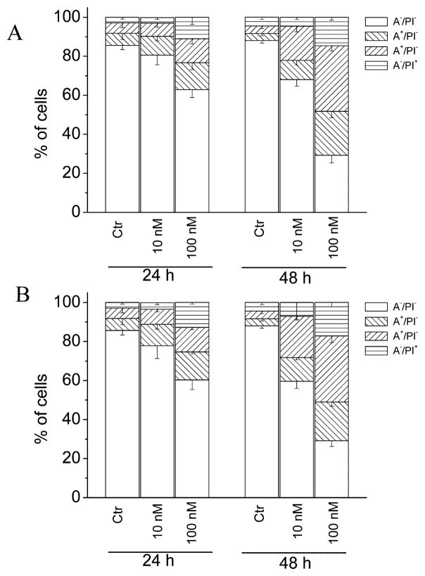
Figure 5.
Flow cytometric analysis of apoptotic cells after treatment of HeLa cells with 4i (A) or 4n (B) at the indicated concentrations after incubation for 24 or 48 h. The cells were harvested and labeled with annexin-V–FITC and PI and analyzed by flow cytometry. Data are represented as the mean ± SEM of three independent experiments.

