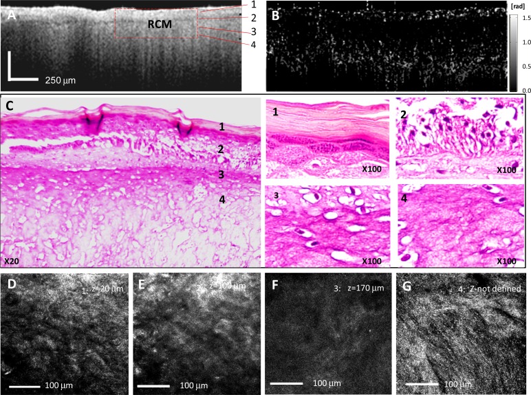
Fig. 7.
Cross-sectional OCT and en face RCM images of the EpiDermFT Skin Model after injury. (A): OCT image. (B): PS OCT image showing very low birefringence of the dermal layer. (C): Histology confirming tissue degradation at all depths; cells are no longer differentiated. (D-G): RCM images showing tissue morphology at different depths. The heat has killed the cells and neither the cobblestone appearance of the corneum layer (D) and the polygonal shape of the epidermal cells (E), or the dermal papillae (F) and collagen fibers in the upper dermis (G) are well distinguishable.

