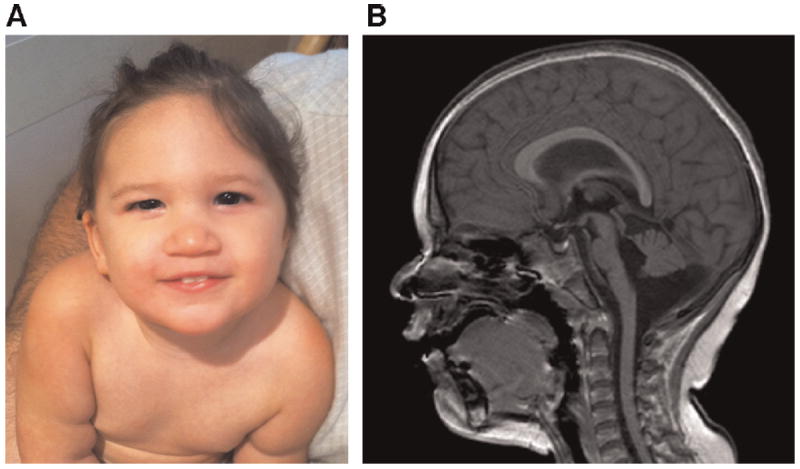
FIG. 1.

A: Patient at age 19 months. B: Sagittal FFE magnetic resonance image demonstrating a thin and abnormally shaped corpus callosum, hypoplasia of the brainstem, hypoplasia of the inferior cerebellar vermis, and a prominent cisterna magna [Color figure can be viewed in the online issue, which is available at www.wileyonlinelibrary.com].
