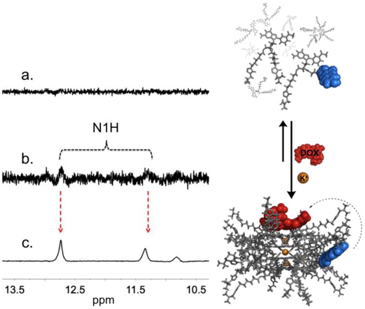
Fig. 2.

Partial 1H NMR spectra (500 MHz, 10% D2O in LiPB, pH 7.4, 298.2 K) of 5 mM sample with: (a) a mixture of 0.06 equiv of 1 and 0.94 equiv of 2; (b) same as (a) but with 0.06 equiv of DOX, where the N1H peaks (corresponding to the formation of a hexadecamer) begin to show; (c) same as b but with 1 M KCl showing a clearly defined double set of N1H signals. The right panel illustrates the equilibrium between (top) monomeric subunits of 1 and 2 and loosely bound aggregates (LBA); and (bottom) one the potential complexes between DOX and the various possible heteromeric supramolecular G-quadruplexes (hSGQ•DOX).
