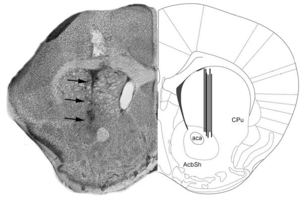
Figure 6. Schematic representation of microdialysis probe locations.

The figure shows a coronal section of the mouse brain through the dorsal striatum (caudate/putamen) region. In the left hemisphere is shown a representative Nissl-stained section revealing the tract associated with the tip of an inserted probe (dark arrows). In the right hemisphere is shown the corresponding region in a stereotaxic atlas [51]. ac = anterior commissure; Acb = Nucleus accumbens of the ventral striatum; CPu = caudate/putamen of the dorsal striatum; LV = Lateral ventricle.
