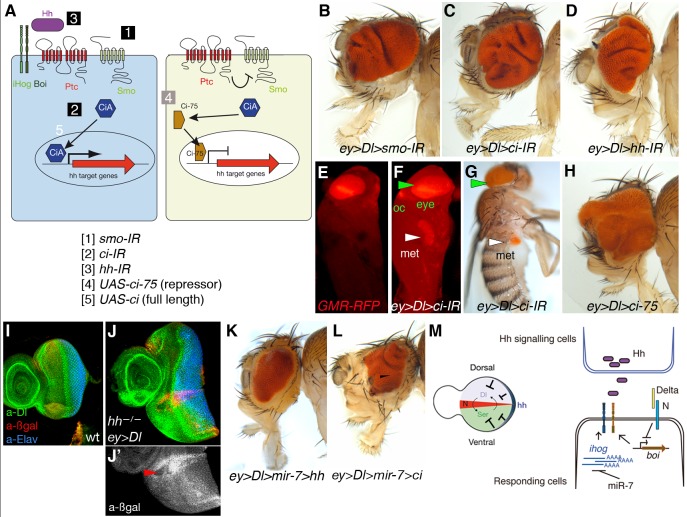
Figure 4. Downregulation of elements in the Hh pathway or overexpression of the repressor form of ci co-operates with Dl overexpression to trigger tumour growth in the Drosophila eye.
(A) Schematic representation of Hh signalling and the UAS transgenes used to downregulate by RNAi (IR) or activate Hh pathway components. (B–D, H, and K–L) Representative adult heads of female flies of combinations of the indicated UAS transgenes and ey-Gal4 are shown. (E–F) Fluorescent images of Drosophila pupae of sibling control (ey>Dl, E) or ey>Dl>ci-IR (F). (G) Adult fly of ey>Dl>ci-IR with a metastatic (met) growth in the abdomen. Eye tissue in the endogenous site (green arrowheads) and distant site (white arrowheads) is labelled by the retinal-specific GMR-myrRFP marker (E, F) or the retinal-specific red pigments (G). (I–J′) Third instar wild type of sized eye disc (I) and ey>Dl eye disc carrying clones of hhAC labelled by the absence of arm-lacZ (ßgal, red in J and grey in J′). Arrowhead points to a clone and its associated twin spot (high red staining). (M) Model of antagonistic interaction between Hh and Notch signalling in normal eye imaginal disc (left) and model of regulatory interactions among the microRNA, Notch pathway, and the Hh receptors ihog and boi (right). Genotype in (J) is: yw ey-Flp; ey-Gal4 UAS-Dl/+; FRT82B hhAC/FRT82B arm-lacZ.

