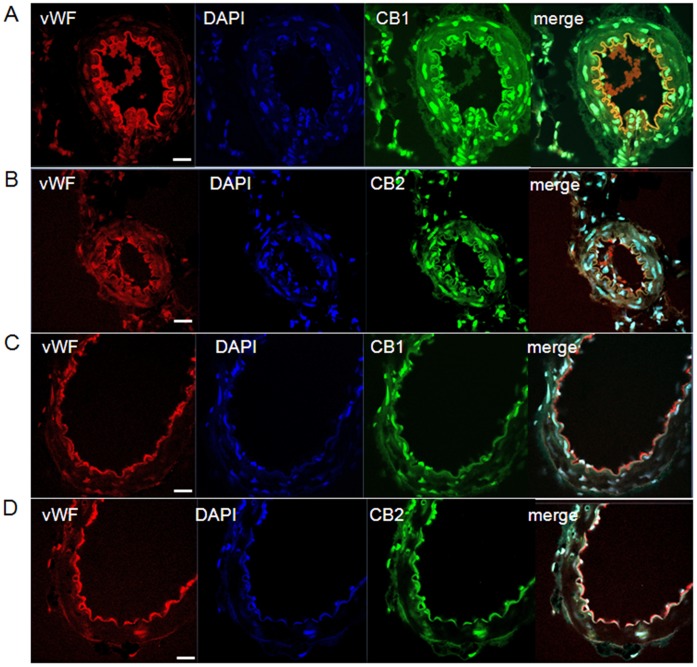
Figure 6. Localization of CB1 and CB2 receptors in mesenteric arteries.
Panels show confocal microscopic images of von Willebrand factor (vWF, red), DAPI (blue), CB1 and CB2 receptors (green) in mesenteric arteries from lean Zucker rats (LZRs, A and B) and obese Zucker rats (OZRs, C and D). Endothelium-intact mesenteric sections were immunolabeled with antibodies against CB1, CB2 and von Willebrand factor. The nuclei were counterstained with DAPI. An overlay of the vWF, DAPI and CB1 or CB2 images is presented (merge). Endothelial localization of CB1 and CB2 receptors is shown in the merged images (yellow). The images are representative of three separated experiments. N = 5/group. Scale bar = 40 µm.

