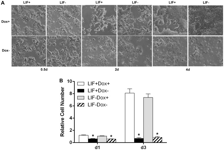
Figure 3. Effects of hKlf4 induction on mES cell growth.
The inducible hKlf4 mES cells were cultured in the presence or absence of LIF. (A) Microscopy was carried out with the Zeiss Axio Observer A1 inverted microscope and an AxioCam MRM camera (Bright field, 5x objective lens). (B) Cells were seeded in triplicate wells at 3×10⁴ cells per well of 24-well plates. After induction for one day and three days, cells were trypsinized and counted with a coulter counter (Beckman Coulter Z1 Particle Counter). The cell numbers were plotted relative to the cell numbers of the LIF+Dox+ at day one. Bars represent the average±SEM of two independent experiments with triplicates each. LIF+Dox− was compared with LIF+Dox+, and LIF−Dox− was compared with LIF−Dox+ for statistical significance. *, p<0.05.

