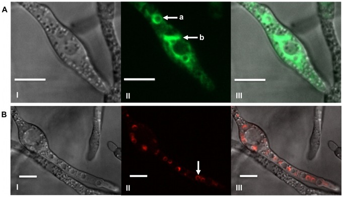
Figure 1. Localization of Tri1p and Tri4p in toxin induced cells.
A) Tri1p::GFP localized to a stationary spherical organelle (a) and a membranous network (b) within the cytoplasm of strain PH-1Tri1::GFPA. Confocal bright field DIC (I); GFP (II); and GFP and DIC (III) overlay images of cells are shown. B) Localization of Tri4p::RFP to a stationary spherical organelle (arrow) within the cytoplasm of strain Tri4::RFPB. Confocal bright field DIC (I); RFP (II); and RFP and DIC overlay images are shown. Both strains were incubated in TBI medium for 36 h. Scale bar = 10 µm.

