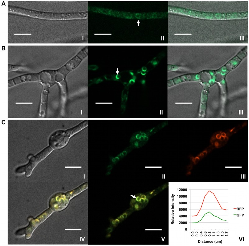
Figure 5. Re-patterning of Hmr1p fluorescence in toxin induced cells.
(A) Localization of Hmr1p::GFP in strain PH-1Hmr1::GFP 36 h after suspension in MM where trichothecene biosynthesis does not occur. Confocal bright field DIC (I); GFP (II); and DIC and GFP (III) overlay images are shown. Hmr1p::GFP fluorescence corresponds mostly to diffuse membranous structures within the cell but occasionally to spherical bodies (arrow). (B) Localization of Hmr1p::GFP in strain PH-1Hmr1::GFP 36 h after suspension in TBI medium. Confocal bright field DIC (I); GFP (II); DIC and GFP (III) overlay images show a shift in localization of Hmr1p::GFP (arrow) primarily to spherical organelles. (C) Co-localization of Hmr1p::GFP and Tri4p::RFP in strain PH-1Hmr1::GFP/Tri4::RFP 36 h after suspension in TBI medium. Confocal bright field DIC (I); GFP (II); RFP (III); and GFP/RFP/DIC (IV) overlay images are shown. Co-fluorescence of GFP and RFP (V) within the periphery of a spherical organelle (arrow) and signal intensity of GFP and RFP emission spectra (VI) generated from a line bisecting the spherical organelle show spatial overlap of the separate emission fluorescence. Scale bar = 10 µm.

