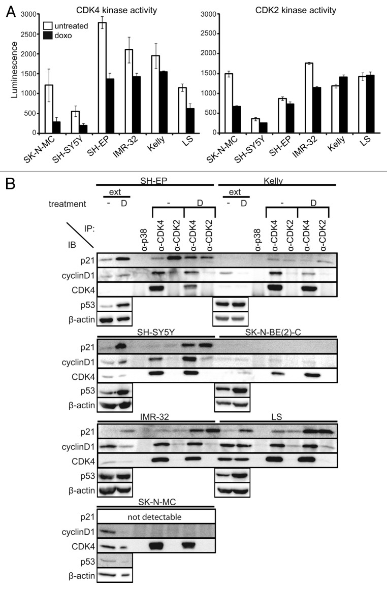
Figure 2. High CDK4 and CDK2 activity after doxorubicin treatment of MYCN-amplified cells. (A) CDK4 and CDK2 activity were analyzed 48 h after treatment using RB and histone 1 as substrates, respectively. Luminescence directly correlates to the amount of produced ADP, indicative for kinase activity. Data are presented as mean ± SD of duplicates. (B) Whole-cell protein extracts were prepared 48 h after doxo treatment and immunoprecipitated with anti-CDK4, anti-CDK2 or control anti-p38 antibodies. Then, 50 µg of whole-cell protein extracts (ext) and 500 µg of the immunoprecipitates (IP) were separated on 12.5% SDS-PAGE. β-actin was used as loading control for whole-cell protein extracts.

An official website of the United States government
Here's how you know
Official websites use .gov
A
.gov website belongs to an official
government organization in the United States.
Secure .gov websites use HTTPS
A lock (
) or https:// means you've safely
connected to the .gov website. Share sensitive
information only on official, secure websites.
