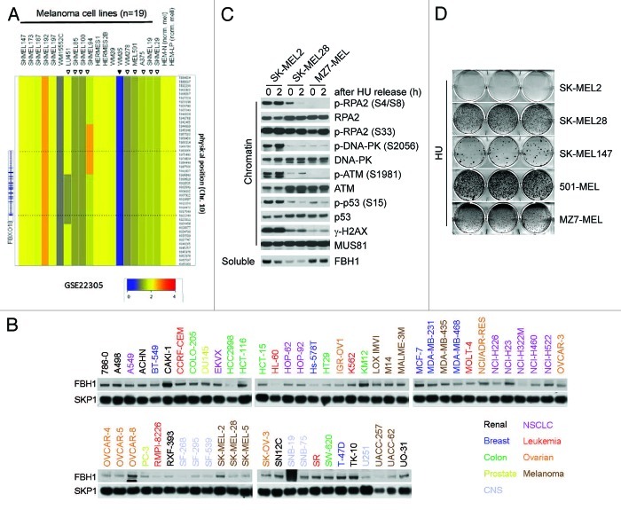
Figure 1. Melanoma cells expressing either low levels of FBH1 or mutated FBH1 display an attenuated DSB signaling response to hydroxyurea. (A) Affymetrix genome-wide human HD-SNP 6.0 array copy number data of the FBH1 locus in 19 melanoma cell lines and in two primary neonatal melanomas is shown. The data are also accessible at the NCBI-GEO (www.ncbi.nlm.nih.gov/geo/) database,4 accession #GSE22305.3 (B) The NCI-60 cell line panel was obtained from the Developmental Therapeutics Program of the National Cancer Institute (http://dtp.cancer.gov/branches/btb/characterizationNCI60.html).5 Whole-cell extracts were subjected to immunoblotting for the indicated proteins. Four melanoma cell lines (SK-MEL-28, SK-MEL-5, UACC-257 and UACC-62), out of the nine in this panel, display low FBH1 protein levels. (C) The indicated melanoma cells were treated with hydroxyurea (HU) for 24 h (time 0) and released for 2 h into fresh medium. After harvesting, cells were fractionated into soluble and chromatin fractions, and lysates were immunoblotted for the indicated proteins. (D) The indicated melanoma cells were treated with hydroxyurea for 72 h, released, cultured for an additional 15–20 d and stained with crystal violet. Images show representative examples.

An official website of the United States government
Here's how you know
Official websites use .gov
A
.gov website belongs to an official
government organization in the United States.
Secure .gov websites use HTTPS
A lock (
) or https:// means you've safely
connected to the .gov website. Share sensitive
information only on official, secure websites.
