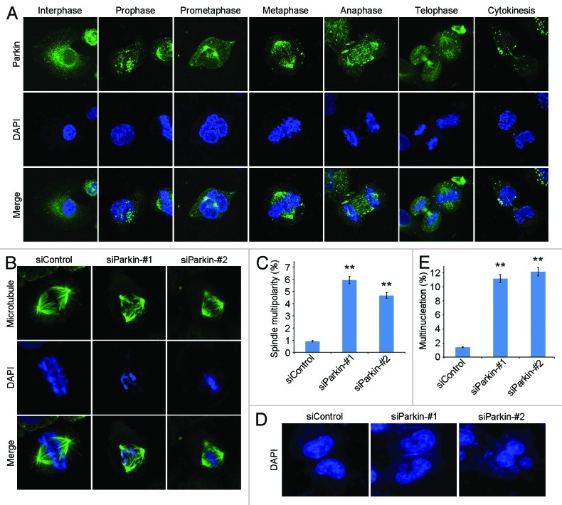
Figure 4. Knockdown of parkin expression increases the frequency of spindle multipolarity and multinucleation. (A) Immunofluorescence microscopic analysis of the subcellular localization of parkin during the cell cycle. Cells were stained with anti-parkin antibody and the DNA dye DAPI. (B) Transfection of parkin siRNAs leads to the formation of multipolar spindles. Cells transfected with control or parkin siRNAs were stained with anti-α-tubulin antibody and DAPI. (C) Experiments were performed as in (B), and the percentage of mitotic cells with multipolar spindles was calculated. (D) Parkin siRNAs result in multinucleation. Cells transfected with control or parkin siRNAs were stained with DAPI. (E) Experiments were performed as in (D), and the percentage of multinucleated cells was determined. ** p < 0.01 vs. control.

An official website of the United States government
Here's how you know
Official websites use .gov
A
.gov website belongs to an official
government organization in the United States.
Secure .gov websites use HTTPS
A lock (
) or https:// means you've safely
connected to the .gov website. Share sensitive
information only on official, secure websites.
