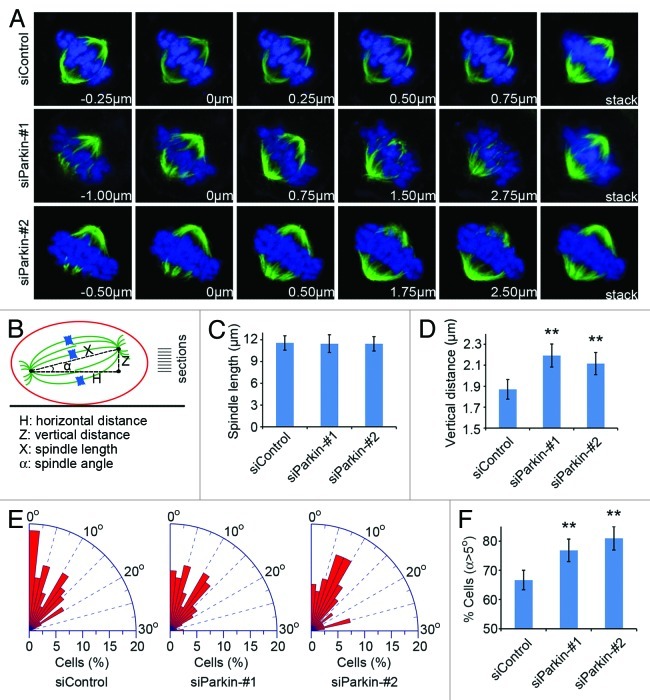
Figure 5. Parkin deficiency leads to spindle misorientation. (A) Cells were transfected with control or parkin siRNAs and stained with anti-α-tubulin antibody (green) and DAPI (blue) and the image series of mitotic cells were shown. The position of the Z stage of the mitotic spindle is indicated in µm, and stack refers to the projected image. (B) Scheme describing the method used for analysis of various parameters of the mitotic spindle. The vertical distance (Z) and the horizontal distance (H) between the two spindle poles were measured directly with the LASAF software. The corrected spindle length (X) was calculated based on the pythagorean theorem. The angle (α) between the spindle axis and the substratum plane was determined with the inverse trigonometric function. (C–E) Experiments were performed as in (A), and the spindle length, the vertical distance between the two spindle poles and the spindle angle were measured as described in (B). (F) Quantification of the percentage of mitotic cells with spindle angles over than 5°. ** p < 0.01 vs. control.

An official website of the United States government
Here's how you know
Official websites use .gov
A
.gov website belongs to an official
government organization in the United States.
Secure .gov websites use HTTPS
A lock (
) or https:// means you've safely
connected to the .gov website. Share sensitive
information only on official, secure websites.
