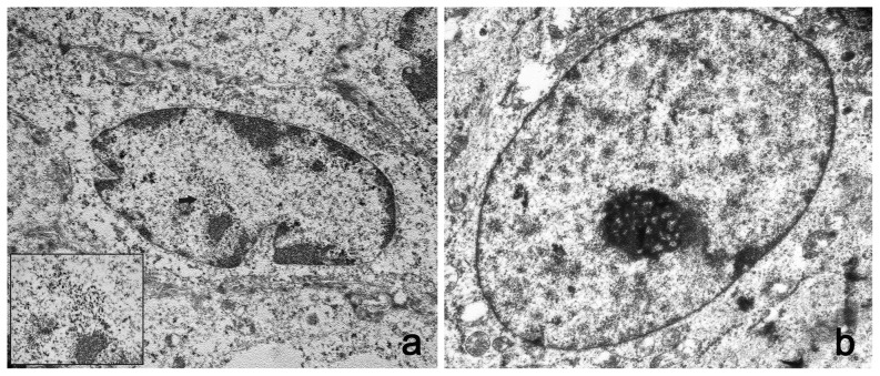
Figure 8. Presence of viral particles in bubaline urothelial cells.
A) Numerous electron dense particles, 45–50 nm in diameter (black arrows) were seen in the nuclei. X 15,000. The size and shape of these particles are consistent with the submicroscopic features of viral particles (insert). X 50,000. B) Normal urothelial cells. No electron dense particles are seen in the nucleus of a normal urothelial cell. X 15,000.

