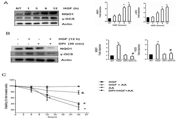
Figure 3.
HGF induces the expression of Nrf2 target genes. Total proteins were prepared from cells treated with 50 ng/ml HGF for 1, 3, 6, and 12 h and subjected to Western blot analysis using indicated antibodies. In parallel experiments, cells were pretreated with 10 μM diphenylene iodonium (DPI) for 30 min before HGF exposure for 12 h. (A) time-dependent increment in NQO1 and γ-GCS. Representative Western blot and densitometry analysis of protein content relative to actin used as loading control. Results are shown as the means ± SEM of 3 experiments. (B) Inhibition of NADPH oxidase abrogates HGF-induced expression of Nrf2 target genes. Representative Western blot and densitometry of protein levels relative to actin used as internal control. Each bar represents the mean ± SEM of at least three independent experiments carried out in triplicate. * P ≤ 0.05 vs not treated cells (NT); # P ≤ 0.05 vs HGF treated cells (HGF). (C) Effect of HGF pretreatment (12 h) on cell viability in cultures treated with antimycin A (AA, 15 μM) for 0, 6, 12, and 24 h. In parallel experiments, cells were pretreated with DPI for 30 min before addition of HGF. Each point represents the mean ± SEM of at least four independent experiments carried out in triplicate. * P ≤ 0.05 vs not treated cells; # P ≤ vs HGF treated cells (12h) and AA (24h); & P ≤ 0.05 vs cells treated with AA (24h).

