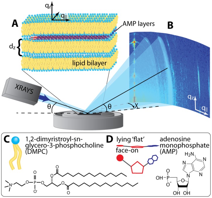
Figure 1. Schematic showing the experimental setup.
A Lipids and AMP form a highly oriented multilamellar structure with the AMP molecules confined between the bilayers. B Diffraction of X-rays from the highly oriented solid supported lipid/AMP complexes. qz and q|| are the out-of-plane and in-plane components of the scattering vector, Q. Chemical structures and representations of C dimyristoylphosphocholine (DMPC) and D adenosine monophosphate (AMP) molecules are depicted.

