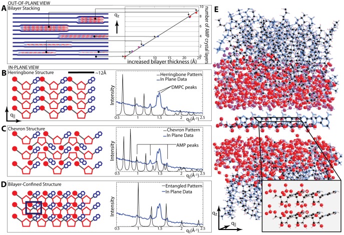
Figure 5. Crystal structure determination.
A Schematic of out-of-plane structure of the lipid/AMP complex. The lamellar spacings determined in Figure 4 all fall on a master curve. The thickness of a single AMP layer, Δd, is determined by the slope to Δd = 2.67 Å. B-D show proposed AMP in-plane crystal structures and the resulting diffraction pattern (black) compared against the peak locations in our in-plane data (blue). B A herringbone structure would be geometrically favorable due to the shape of AMP. The diffraction pattern, however, does not agree with our experimental data. C The chevron pattern is also a favorable structure for the packing of ’v’ shaped molecules, however the diffraction pattern that would be produced from this structure is not consistent with the experiential data. D The tetragonal 2-dimensional unit cell with lattice parameters of a = 6.25 Å and b = 4.8 Å gives the most plausible structure of the AMP molecules. E Molecular representation of the crystalline AMP between the stacked DMPC bilayers; in-plane representation below. The molecular coordinates for the DMPC bilayer were taken from [29]. Molecular structure files of the structures in D and E are provided in Structure Files S1 and S2.

