Fig. 4.
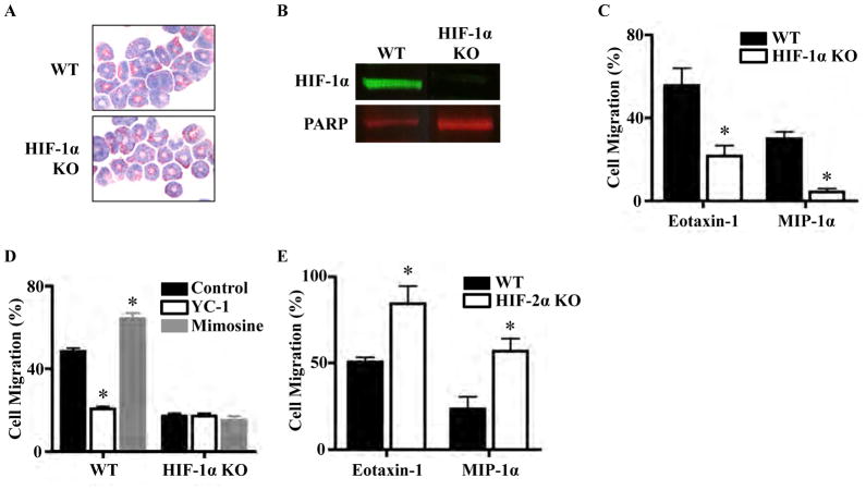
HIF-1α and HIF-2α regulate eosinophil chemotaxis. a Giemsa–Wright stain of C57BL/6 WT and HIF-1α KO eosinophils. b HIF-1α KO eosinophils have much decreased HIF-1α levels com- pared to WT eosinophils by Western blot, with PARP as a nuclear housekeeping protein control. c Transwell chemotaxis assay demon- strating that HIF-1α KO eosinophils migrate less than WT toward eotaxin-1 and MIP-1α (*P < 0.001). d HIF-1α antagonist YC-1 and agonist mimosine alter migration of WT but not HIF-1α KO eosino- phils (*P < 0.001). e HIF-2α KO eosinophils migrate more than WT toward eotaxin-1 and MIP-1α (*P < 0.01). Each condition was done in duplicate during the experiment, and each experiment was repeated three times

