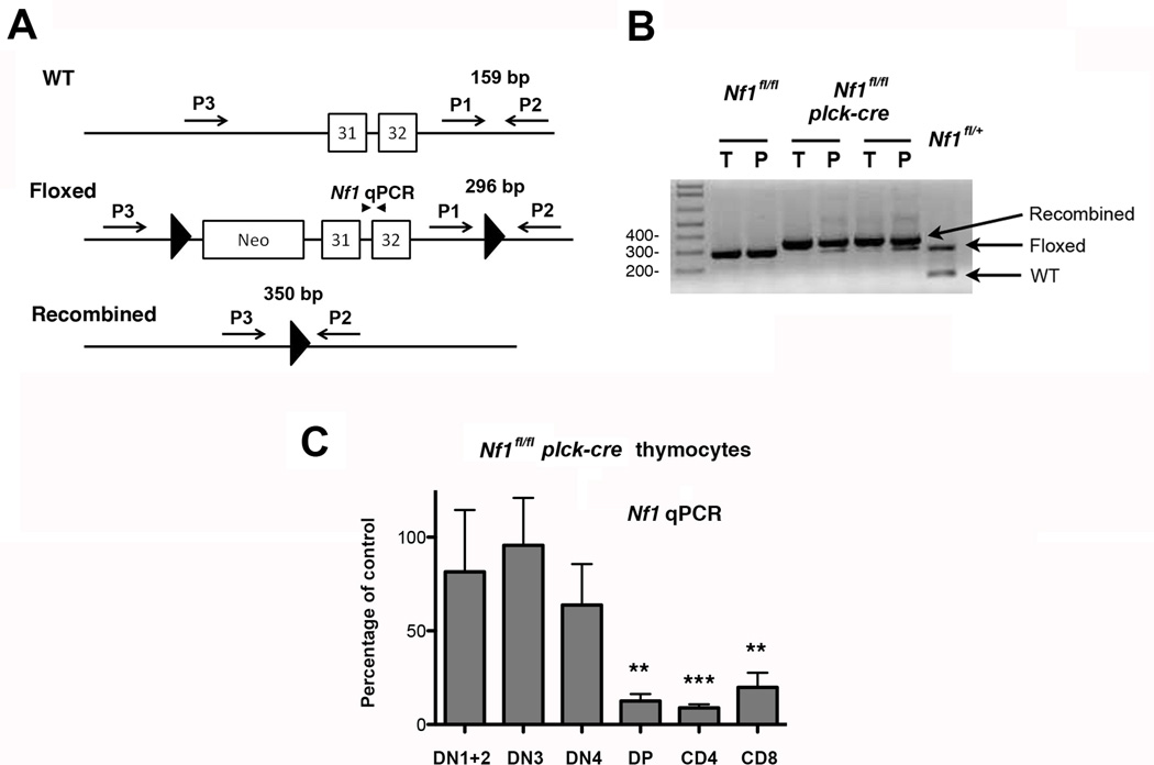
Fig. 1.
T cell-specific deletion of NF1. A, Shown is part of the wild type (WT), floxed and recombined Nf1 locus and position of primers P1-P3 (arrows) and qPCR primers (arrowheads) used to determine the extent of NF1 deletion in Nf1fl/fl plck-cre mice. B, Genomic DNA was extracted from whole thymus (T) and purified peripheral splenic and LN T cells (P) from Nf1fl/fl and Nf1fl/fl plck-cre mice and analyzed by PCR using primers P1-P3 in a single reaction. Shown are the results of a representative analysis performed with two Nf1fl/fl plck-cre mice and a littermate Nf1fl/fl control. For comparison, the same PCR reaction was performed upon tail genomic DNA from a Nf1fl/+ mouse. C, Genomic DNA was prepared from the indicated sorted thymocyte subpopulations from littermate Nf1fl/fl and Nf1fl/fl plck-cre mice and relative Nf1 gene amounts were determined by qPCR using primers shown in A. For each subpopulation, the percentage amount of Nf1 in Nf1fl/fl plck-cre mice compared to Nf1fl/fl mice was calculated. Experiments were performed with three independent pairs of mice. Shown is the mean percentage of Nf1 expression + 1 SEM in Nf1fl/fl plck-cre mice compared to Nf1fl/fl mice. Statistical significance was determined using a Student one-sample t test. **p <0.01, ***p <0.001.

