Figure 4.
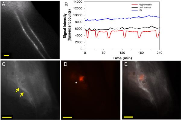
Dysfunction of popliteal collecting lymphatic vessels in a B16 footpad tumor bearing mouse with LN metastases. (A) Screen capture of video beginning five minutes after L-ICG injection (5 μL of 30 μM) into hind paw. (B) Quantification of afferent popliteal collecting lymphatic pulsing and LN fluorescent signal demonstrating strong pulses in right afferent vessel with irregular pulses in left afferent vessel. (C) NIR image of popliteal LN after skin removal showing intense L-ICG signal at junction of left afferent vessel and LN (yellow arrows). (D) Fluorescent image demonstrating strong tdTomato signal on left side of popliteal LN (white asterisk). (E) Overlay of images demonstrating strong tdTomato signal in LN at site of afferent vessel entry into popliteal LN. Scale bars: 500 μm.

