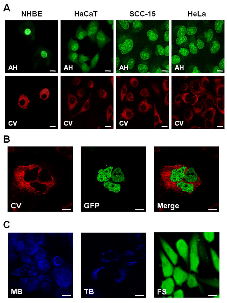
Figure 1. In vitro contrast agent testing.
(A) Comparative confocal images of cultured normal (NHBE) and cancer cells (HaCaT, SCC-15, HeLa) from left to right, topically stained with Acriflavine Hydrochloride (AH; upper panel) or Cresyl Violet (CV; lower panel). (B) Cultured H2B-GFP expressing cells stained with CV: left, CV (red fluorescence) only; middle, GFP (green fluorescence); right, co-localization of CV and GFP. (C) Cultured cells topically stained with Methylene Blue (MB; left), Toluidine Blue (TB; middle) and Fluorescein Sodium (FS, right). The common scale bar is 20 μm wide.

