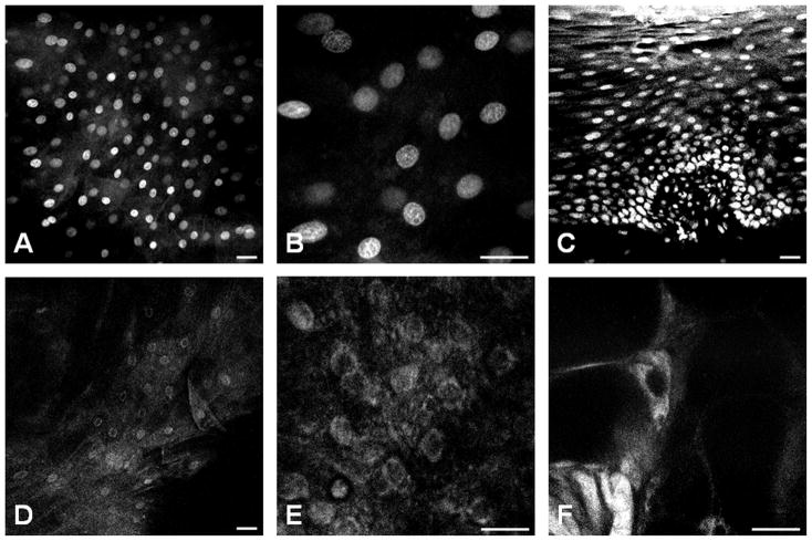
Figure 2. Ex vivo contrast agent testing.
Confocal imaging comparing the nuclear morphology of the oral epithelium after topical application of Acriflavine Hydrochloride (A–C) or Cresyl Violet (D–F). A & B, an en face image taken from a normal epithelium at 50 μm from the surface and B is from the same field of view with magnified A. C, an on edge transversal section of oral mucosa showed multilayering and uniform polarity of differentiated cells that distinguish the stratified squamous epithelium from the basal cell layer (closely packed low cuboidal nuclei at the bottom), the spinous cell layer (evenly spaced out, slightly ovoid shaped nuclei with their long axis parallel to the surface), and the parakeratinized superficial layer (tightly packed with smaller, elongated nuclei at the top). D & E, an en face image taken from a normal epithelium at 50 μm from the surface show fainter cytoplasmic and perinuclear uptake of CV (E is the same field of view with magnified D). F, a star-like cell with elongated cytoplasmic protrusion shows bright intra-cytoplasmic fluorescence. This cell, according to its morphology and intraepithelial location, is consistent with a Langerhans (or antigen presenting) cell. The common scale bar is 50 μm wide.

