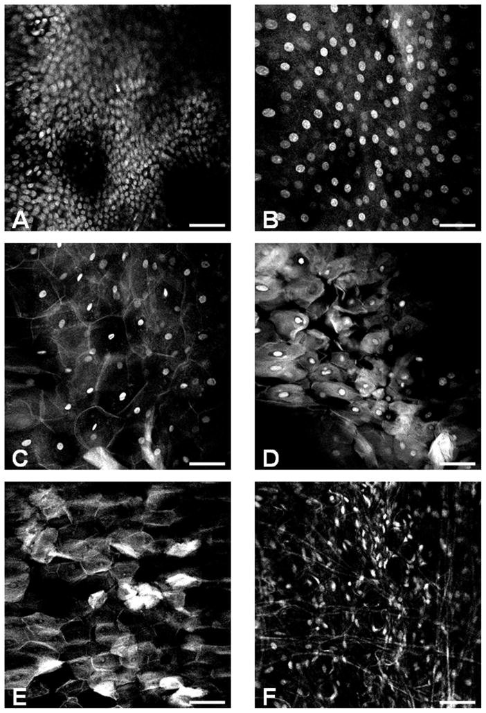
Figure 3. Representative confocal imaging histology of normal oral mucosa.
An atlas of the oral mucosa displaying different layers of the stratified squamous epithelium and subepithelial connective tissue: (A) basal cell layer, (B) parabasal cell layer (50 μm above A), (C) spinous cell layer, (D–E) superficial parakeratinized or keratinized layer, (F) Subepithelial connective tissue. The scale bar is 100 μm wide.

