
Figure 1.
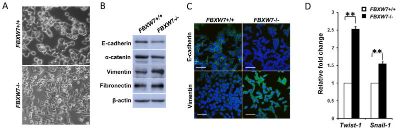
Depletion of FBXW7 in HCT116 cells leads to Epithelial-mesenchymal transition. (A) Morphology comparison of HCT116 FBXW7−/− and its control cells under bright field. (B) Western blot analysis of indicated EMT markers in HCT116 FBXW7−/− and its control cells. (C) Immunofluorescence staining for indicated EMT markers (Green) in HCT116 FBXW7−/− and its control cells. DAPI staining is blue. Scale bar is 20μm. (D) qRT-PCR analysis of Twist-1 and Snail-1 expression in HCT116 FBXW7−/− and its control cells. Data were presented as means ± Standard deviation from three independent experiments in triplicates. **P <0.01 was obtained from Student’s t-test.