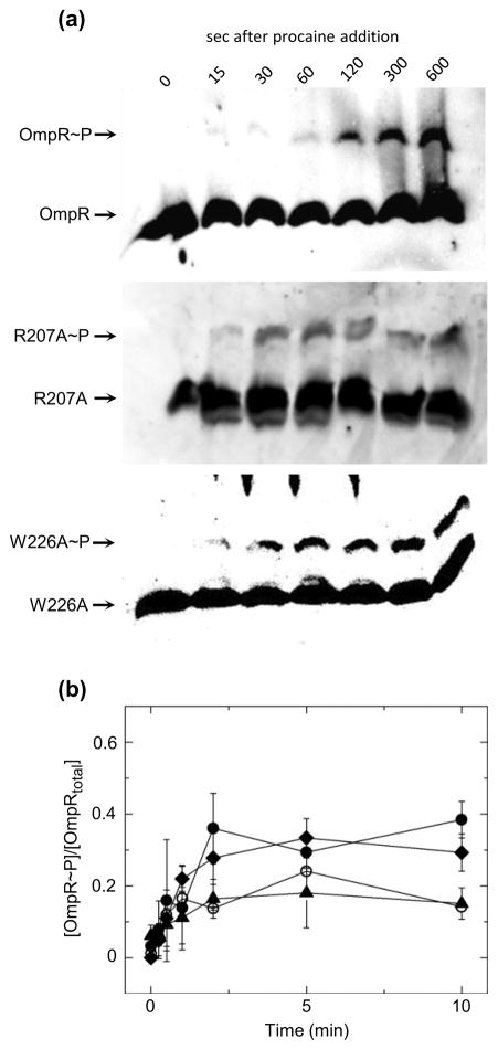
Fig. 5.
Time-dependent change in plasmid-expressed OmpR, R207A and W226A phosphorylation in ΔompR cells following treatment with procaine. (a) Representative western blots of phosphoprotein affinity gel electrophoretic separation of ΔompR cells expressing either OmpR, R207A or W226A following treatment with procaine. Lane 1 in all blots contains untreated cells lysates and lanes 2–7 contain cell lysates at indicated times following treatment with procaine. (b)Quantitation of the extent of phosphorylation of OmpR (circles), R207A(triangles) or W226A (diamonds) following treatment with procaine. Points reflect the mean values of 3 separate experiments, with error bars reflecting the standard deviation from the mean.

